Category: Treatments

25 August 2020 by rommer 0 Comments

Aquatherapy / Hydrotherapy

HYDROTHERAPY (ACUATIC REHABILITATION)

What is Hydrotherapy?

Hydrotherapy (pool therapy) is a method of treating diseases and functional losses by taking advantage of the physical properties of water. Water is one of the oldest methods used for physical therapy.  The heat and hydrostatic properties of the water are utilized. With the help of the buoyancy of water, the load carried by the body is reduced, thus it is possible to perform movements that cannot be done normally or that cause a lot of pain while doing it.


What are the benefits of hydrotherapy?

  • It plays an important role in reducing pain and sensitivity
  • It increases joint movements and helps muscles relax.
  • It helps to develop respiratory muscles and the body starts to use oxygen more efficiently.
  • It increases muscle strength and endurance by using the resistance property of water
  • Difficult and tiring exercises that cannot be done outside of the water become simple and fun, and the self-confidence and motivation of the patient who can perform the movements more easily increases.
  • Due to the less effect of gravity in water, it accelerates healing and the healing process of some diseases.
  • It prevents excessive load on the person’s muscles and movements are made easier.
  • It helps to activate the lymph system.
  • It helps to control edema.


Hydrotherapy applications in neurological diseases

In patients with spinal cord injuries, standing and walking activities and some exercises may cause excessive load on the joints and injuries due to the lack of muscle strength. These exercises can be done more comfortably and safely by using the buoyancy of the water. Again, thanks to the buoyancy of the water, standing and walking activities that are hard on dry ground can be done more easily and it is beneficial in increasing muscle strength.

In diseases such as hemiplegia (stroke), MS (Multiple Sclerosis), activities performed with difficulty on dry ground due to contractions can be performed more easily with the relaxation effect of the muscles provided in water.

Hydrotherapy applications in orthopedic diseases

After orthopedic surgeries, especially in cases where it is inconvenient to give weight to the joint immediately, hydrotherapy applications can be used because the load on the joint is less. Since the exercises performed outside the water during rehabilitation can be challenging for the person, more comfortable exercise can be done by taking advantage of the buoyancy effect of the water in the pool.


What are the main diseases for hydrotherapy?

  • Cerebral Palsy
  • Multiple Sclerosis (MS)
  • Muscular Dystrophy,
  • Guillan Barre Syndrome,
  • Musculoskeletal System Injuries,
  • Stroke – paralysis
  • Spinal Cord Injuries
  • Joint Injuries,
  • After Fractures and Surgical Procedures,
  • Fibromyalgia
  • Rheumatic Diseases
  • Sports Injuries
  • Soft Tissue Injuries,
  • Obesity
  • Osteoarthritis (Calcification)
  • Balance Problems,
  • Polio

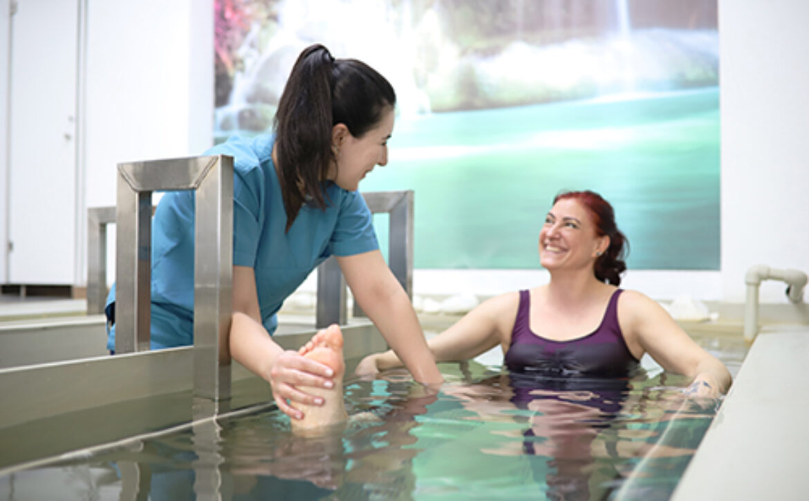
How is hydrotherapy applied in ROMMER?

In our center, the hydrotherapy application is personally planned after the examination of the specialist physician and applied individually in the presence of a physiotherapist. Since thermal water is used in the pool in our center, the effect of these healing waters is also benefited. Hydrotherapy application is very beneficial for mobilization in diseases such as cerebral palsy and stroke (paralysis) and contributes to the success of rehabilitation.

25 August 2020 by rommer 0 Comments

Meniscal Tears

MENISCAL TEARS

What is Meniscal Tear?

The menisci in the knee joint are cartilage parts in the shape of a half moon. There are 2 meniscus in the knee joint, internal and external. The menisci are located between the tibia and the thigh. Menisci are structures with dense collagen fibers to resist compression, elasticity and therefore have important functions. The menisci absorb the incoming shocks, distribute the load over a wider area and protect the articular cartilages from high pressure and help the stability of the knee.

How Does a Meniscus Tear Occur?

Meniscus tears can occur in people of all ages, although meniscus tears can be seen as an athlete’s disease because it can be seen frequently in athletes. Injuries are mostly seen during sudden rotational movements. Since the meniscus tissue is solid in young individuals, severe trauma must be present in order for meniscus tears to be seen. However, due to the degeneration that occurs in the meniscus at advanced ages, these individuals can have tears even in mild traumas.

What Are Meniscal Tear Symptoms?

The most important clinical finding of meniscus tears is pain. Pain usually occurs during the knee-bending motion. However, depending on the size of the tear, there may also be complaints of locking and snagging in the knee. As there will be compression in the meniscus during stair use and squatting, pain may increase and locking may occur in the bent knee.

There may also be fluid increase in the joint space in the knee where meniscus tears occur. Patients describe this as a feeling of swelling or fullness in the knee. Early diagnosis is important in meniscus tears. As a result of the meniscus being torn for a long time, stability problems may occur in the knee, and accordingly calcification and cartilage damage may occur.

What To Do In Physical Therapy For Meniscus Tears?

Physical therapy methods are used as conservative treatment in incomplete meniscus tears. The purpose of the treatment of these patients is the elimination of complaints such as pain, swelling and joint restrictions and strengthening of the muscles around the knee.

Physiotherapy plays an important role in edema control, prevention of muscle atrophy, pain control and returning the patient to normal daily life activities.


Why Is Physiological Treatment Important After Surgery For Meniscal Tears?

Advanced meniscus tears are now treated surgically. Depending on the meniscus tear, surgery is performed in two ways. Depending on the location and condition of the tear, surgery is performed either as meniscus repair or meniscectomy (removal of the meniscus).

After the surgery, the role of physiotherapy is great. Depending on the type of surgery performed, the duration of physical therapy also varies between 2 weeks and 6 weeks. The main purpose of physical therapy after surgery is reducing pain and inflammation, increasing muscle strength and endurance, helping the joint regain function as the articular cartilage has to adapt to maintain function without  meniscus .

For this purpose, physical therapy modalities and appropriate exercise programs are used. Since the exercises to be performed are determined gradually in the postoperative period and are planned according to the patient’s condition, the exercises should not be applied unconsciously.

25 August 2020 by rommer 0 Comments

Orthopedic Rehabilitation

After orthopedic surgeries, plaster applications, splint applications, prosthesis applications, stiffness and rigidity usually occur in the joints of the patients. After the plaster or splint treatment, the patient cannot use that joint as soft and comfortable as before. The reason for this is the inactivity experienced in the muscles due to the joint. In addition to complaints such as swelling in the joint, limitation of movement and pain with movement, a significant weakness will be observed in the muscles. At this point, orthopedic rehabilitation will come into play.

What is Orthopedic Rehabilitation?

It is a sub-unit specialized in orthopedic rehabilitation, physical therapy and rehabilitation, and looks after a very intense disease group. After the patient, who is suitable for orthopedic rehabilitation, is evaluated by a physical therapist, a special rehabilitation program is prepared for the patient and in this process, the patient and the physiotherapist start to work one to one. The duration of the treatment and the number of sessions to be performed vary according to the patient’s problems.

Orthopedic Rehabilitation Applies to Which Patients?

Orthopedic rehabilitation is applied in all orthopedic cases of the musculoskeletal system that require conservative or surgical treatment. Before and after orthopedic intervention (surgery, plaster, splint, prosthesis, etc.), the patient should be evaluated and a short, medium and long-term physical therapy plan should be prepared. Plans for the future of the patient should be discussed with orthopedics and physical therapists and assistants, and an orthopedic rehabilitation protocol should be established at first.

The task is not only to bring the patient to the highest level of functional well-being and rehabilitate it, but also to protect the patient’s current condition and the surgery performed. Orthopedic rehabilitation;

  • Joint stiffness and circulatory disorders after fracture / dislocation
  • Before and after meniscus, ligament injuries and other sports injuries surgeries,
  • Scoliosis and spinal curvature,
  • After flat feet, foot problems and sports injuries,
  • Before and after orthopedic surgeries such as prosthetics. Orthopedic rehabilitation is applied in many diseases that cannot be counted.

What are the Objectives of Orthopedic Rehabilitation?

The aims of orthopedic rehabilitation are to restore or improve the body functions lost due to defects in the patient’s skeletal structure. The aim is to bring the patient back to his / her job and profession, to bring the patient back to his / her job and profession, to make him / her live without needing anyone else as a result of the treatment.

The patient to be treated for orthopedic rehabilitation is prepared for a special rehabilitation program after being evaluated by the physiotherapy and rehabilitation specialist and physiotherapist, taking the opinion of the orthopedic specialist. The rehabilitation process is determined as a result of one-to-one work with physiotherapists according to the patient’s potential, the patient’s contribution to the treatment and compliance.

In Which Diseases Is Orthopedic Rehabilitation Applied Most Frequently?

  • Amputation rehabilitation; It is the surgical removal of some or all or both together with the bone of the arm or leg. 85% of amputations that can be applied due to many reasons are in the legs. After the patients are stabilized after amputation, orthopedic rehabilitation approach should be initiated and stump rehabilitation should be started. Prosthesis and orthosis are applied for the lost area. Rehabilitation continues depending on the location and condition of the prostheses.
  • Endoprosthesis surgeries; Joint prostheses are mostly applied to large joints such as knees, hips and shoulders. Efforts are made not to add new postoperative problems to existing problems already before the surgery. Orthopedic rehabilitation should be started as soon as the patient is stabilized.
  • Fracture rehabilitation; Depending on the condition of closed or operated fractures, physical therapy applications that reduce edema and pain and increase blood flow are performed during the recovery period. After the cast is removed or after the surgical operation, walking sticks or crutches, walking balance training should be given. Orthopedic rehabilitation should be started at the same time.
  • Scoliosis rehabilitation; Physical therapy and rehabilitation are of great importance in the pre- and post-operative period.
  • Cruciate ligament injuries to the knee area.
  • Meniscus damage and tears in the knee.
  • Many diseases such as muscle and tendon injuries due to trauma in the shoulders and elbows are included in the fields of orthopedic rehabilitation.

All of these tables are covered within the orthopedic rehabilitation program. Orthopedic rehabilitation is a team work. The team should include personnel from different disciplines such as orthopedic specialists, FTR specialists, physiotherapists, nurses, psychologists, occupational therapists, orthotics and prosthetics specialists.

25 August 2020 by rommer 0 Comments

Rehabilitaion in Anterior Cruciate Ligament Injuries

REHABILITATION IN ANTERIOR CRUCIATE LIGAMENT INJURIES

What is Anterior Cruciate Ligament Injury?

The Anterior cruciate ligament is an important ligament structure in the knee joint. It prevents the tibia from shifting anteriorly under the femur (thigh bone). It prevents tibial internal rotation. It protects the joint against inward and outward strains.

The anterior cruciate ligament is the one with the least elasticity of the ligaments around the knee. Therefore, the anterior cruciate ligament is the most injured ligament in the knee. Pain, swelling and inflammation symptoms that occur after anterior cruciate ligament injury lead to loss of strength in the muscles, which causes instability in the knee.

What are the Causes of Anterior Cruciate Ligament Injury?

Stress on the knee as a result of sudden turning, stopping or changing direction can cause cruciate ligament and anterior cruciate ligament injuries. Unstable falling after jumping, blows towards the knee can also cause injury.

The use of unsuitable equipment during sports such as football and skiing can also be caused by playing football on unsuitable grounds such as artificial turf.

Treatment with Rehabilitation in Anterior Cruciate Ligament Injuries

In the treatment of anterior cruciate ligament injury, rehabilitation should be applied with a conservative approach, and surgical consultation should be requested when necessary.

Rehabilitation and conservative treatment are decided according to the patient’s age, activity level, profession and future sports-related expectations. The aim of conservative therapy is to return the range of motion in the knee to its normal level, to restore muscle strength, to increase stability and to restore functions to pre-injury levels.

Rehabilitation treatment consists of acute, subacute and chronic phases. The rehabilitation and exercise program to be applied during these periods should be planned individually. Individuals and athletes who want to exercise at a high level in their future life should be evaluated separately according to the degree of damage to the ligament.

Post-Surgical Rehabilitation in Anterior Cruciate Ligament Injuries

After the anterior cruciate ligament operations, the goal is that the patient can continue his / her life without pain and with almost normal joint mobility and muscle strength.

After current anterior cruciate ligament injuries, there are differences in the post operative rehabilitation process according to the graft type preferred in surgery.

After anterior cruciate ligament injury, in the rehabilitation process, the exercises to be done every week are planned according to the condition of the patient, adhering to a certain protocol, and the variety and intensity of the exercises are progressively increased by following the progression.

What are we doing in ROMMER for anterior cruciate ligament injury rehabilitation?

The protocols to be applied after the detailed examination of our specialist physicians to the patients who have undergone an operation or not, who applied to our center after anterior cruciate ligament injuries, are determined. Thanks to this personalized program, it is possible to achieve success in patients with anterior cruciate ligament injury .

25 August 2020 by rommer 0 Comments

Spinal Curvature (Scoliosis)

SPINAL CURVATURE (SCOLIOSIS)

Spinal Curvature (Scoliosis) is a spinal disorder that is often diagnosed by mothers who hurry to bring their children to the doctor.

Our spine is like a bridge that carries about 3/2 of its body weight and is made of 33 vertebrae. When we look at a normal human spine from the side, mild lordosis in the waist and neck region and four curvatures called mild kyphosis in the back and hip area are detected. When viewed from the front, there is no curvature in the spine and it is expected to show a flat structure.

What is Spinal Curvature?

The curvature of the back and lumbar region of the spine (to the right or left) is called scoliosis. Its incidence is about 2-4%. It is seen 8-10 times more in girls than in boys. In only 10% of people with spinal curvature, the curvature progresses to a level that requires treatment.

Scoliosis causes can be different. Scoliosis, which we frequently encounter in our daily life, are mostly idiopathic scoliosis and congenital scoliosis that occur in the 10s and whose cause is not fully determined. Here we will deal mostly with idiopathic scoliosis.

In scoliosis, the curvature of the spine can occur as a single curve in the shape of the letter “C” or as two curves in the shape of the letter “S”. Although ” S ” shaped spinal curvature is seen at all ages, it may not be diagnosed until adolescence. Often it is the mother who first notices the Curvature.

What Are the Types of Spinal Curvature?

Spinal curvatures can occur due to many different reasons. However, the cause cannot always be determined. The most common type is idiopathic scoliosis of unknown cause.

  • Congenital scoliosis (congenital) is usually due to a defect in the spine or fused ribs It progresses very quickly.
  • Idiopathic scoliosis; this group comprises more than 80% of scoliosis cases. It is the most common form of scoliosis. It is called infantile onset if it starts in the 0-3 age group, juvenile onset if it occurs between the ages of 4-9, and adolescent-onset idiopathic scoliosis if it occurs between the ages of 10-18.
  • Early onset scoliosis is a type of scoliosis that usually occurs under the age of 10.
  • Neuromuscular scoliosis occurs as a result of muscle and nerve diseases such as cerebral palsy, polio, cerebral palsy or muscle wasting.

What are the Symptoms of Spinal Curvature?

  • One shoulder to be forward, higher and forward,
  • Shoulders and hips not being aligned,
  • When viewed from behind, one of the shoulder blades is more prominent and protruding,
  • One leg appears to be longer than the other,
  • The trunk and rib cage appear to have slipped to one side,
  • When the person leans forward, the ribs on one side of the body remain higher than the other,
  • Asymmetry in the waist or back of the person.

It is not always easy to predict how scoliosis will progress. However, the period in which the current spinal curvature can progress most rapidly is the first 5 years of life and the rapid increase in height during adolescence. If the curvature at the time of diagnosis is high in the curvatures detected in these periods, the rate of progression will be higher than a low grade curvature. Especially in idiopathic scoliosis, when determining the treatment plan, the age of the child should come first among the criteria considered. The degree of curvature is evaluated in the second place. Faster progression can be expected in girls in terms of gender.

The younger the child’s age when diagnosed, the greater the curvature of the spine will grow and progress. The progression of the Curvature in adult individuals who have completed skeletal development is not as pronounced and rapid at this level. For these reasons, spinal curvature should be monitored very closely in children who are diagnosed with scoliosis at young ages and especially in the rapid growth period of adolescence.

How Is Spinal Curvature Diagnosed?

Early diagnosis of scoliosis is very important as it directly affects the success of the treatment. In addition to a good medical history and physical examination, imaging methods involving the whole spine are required for the diagnosis of scoliosis. The curvature angle should be measured on the anterior, posterior and lateral radiographs of the entire spine while standing. This angle that measures the degree of scoliosis is called the Cobb angle. A method called scoliometer is also used for diagnosis.

The important thing in scoliosis is to determine the degree of rotation accompanying the curvature as well as the curvature. Here, the scoliometer shows us this rotation different from the angle of scoliosis. In addition to plain films, methods such as MRI and tomography are also used in diagnosis and differential diagnosis.

How Should Spinal Curvature Be Treated?

In order to decide on scoliosis treatment, many factors such as the degree of curvature, age of the patient, type of scoliosis, etc. are taken into consideration. Since early treatment affects the results positively, early diagnosis is very important.

In this direction, scoliosis treatment should be completely personalized. For example; the treatment of an 8-year-old child with 30 degrees of Curvature will be different than the treatment of a 18-year-old with 30 degrees of Curvature. Because the younger the child’s age, the greater the potential for the Curvature to progress.

The aim of the treatment is to live a balanced and painless life with an aesthetically smooth spine. General treatment is examined in 3 main categories: observation and follow-up, corset treatment and surgery.

  • Observation and monitoring; It includes the control of spinal curvature at 4-6 month intervals and is the first treatment option. For patients with curvatures less than 20 degrees, whose skeletal development is close to completion, only follow-up and periodic controls will be sufficient. During this follow-up, the patient should include exercise in his / her daily life and increase his / her general body condition.
  • Corset treatment; the aim is to avoid the curvature. It is an effective treatment method for people whose curvature is between 20-40 degrees and continues to grow. However, since corset application is not possible in very young children, general correction and body casts are used. In order for the corset to be effective, the person should apply this treatment for long periods such as 22-23 hours a day. The biggest difference between congenital and idiopathic scoliosis is that corset treatment is not effective in congenital scoliosis.
  • Surgery; If the curvature is above 40-45 degrees in scoliosis patients and especially if the growth continues in children, surgical treatment will be inevitable. Correction and fusion (fixation) operations are applied. Implants such as screws and rods are placed on the back and waist.

What Happens If Spinal Curvature Is Not Treated?

Looking at scoliosis treatment just as a correction of a spine Curvature is an incomplete perspective. Because deformities in the spine, when the degree of curvature reaches a certain level (80-100 degrees), will cause deformities in the ribs and rib cage, and will narrow the chest volume. Especially in growing children, this situation will negatively affect lung development and cause respiratory failure. In other words, scoliosis treatment will not only correct the deformity in the spine, but also prevent possible chest deformities, and consequently, preserve lung functions. For this reason, this should be taken into consideration when making an early surgery decision, especially in young children.

25 August 2020 by rommer 0 Comments

Hemiplegia – Stroke Rehabilitation

HEMIPLEGIA (STROKE / PARALYSIS) REHABILITATION

What is hemiplegia (stroke / paralysis)?

Table known as hemiplegia or stroke / paralysis; is a syndrome that develops as a result of changes in the cerebral circulation and is characterized by dysfunctions in one half of the body.

What are the causes of hemiplegia (stroke / paralysis)?

Cerebrovascular events may occur due to causes such as tumors, inflammation of the vessels, connective tissue and vascular diseases. The most common causes of hemiplegia tables are brain hemorrhage or impaired blood supply to the brain.

What are the disorders seen in hemiplegic patients?

Although hemiplegia is basically defined as the loss of function in one side of the body, it refers to all clinical findings that occur. These clinical findings are also combined with symptoms such as speech disorder, balance disorder, shoulder problems, visual disturbances, urinary and fecal incontinence, memory problems and sensory loss. The findings appear in different ways depending on the affected vessels of the brain and the way they are affected.

In hemiplegic patients, there may be impairments in perception, motor and sensory functions, speech and mental abilities.

What is done in hemiplegia rehabilitation?

The aim of hemiplegia rehabilitation is to try to gain the patient’s physical, social and economic independence and to maximize their functions. Rehabilitation organized for this purpose is applied by a team of physiotherapists, physiotherapists and occupational therapists.

The return of movements after a cerebrovascular event is closely related to the cause, severity and location of the event. The factors affecting the development of the patient are age, motivation, family and socio-economic level of the patient, specific neurological disorders. Therefore, it is very important to start hemiplegia rehabilitation early.

In hemiplegia rehabilitation;

Electrotherapy methods are applied as well as personal neurophysiological exercise approaches.

Technology is also used in hemiplegia rehabilitation. With robotic rehabilitation systems, correct gait training is given to the patient, while computerized balance systems play an important role in increasing the balance and coordination of the person.

What is done in occupational therapy in hemiplegia rehabilitation?

Ergotherapy, on the other hand, enables the person to adapt to daily life, while the sensory integration training also helps the person gain their functions.

People with brain lesions have difficulty in structuring and organizing information. The patient may not be competent in terms of planning, automatic attention and adaptation to the steps required by the job. Therefore, it is important to evaluate and improve sensory perception functions in people with brain lesions. At this point, again, ergotherapy comes into play.

Hemiplegia patients are treated in the best physical therapy and rehabilitation clinics and reintegrated into society and they make more improvements in daily living activities. Hemiplegia rehabilitation in ROMMER Physical Therapy and Rehabilitation Center is successfully applied by experienced physiotherapists and occupational therapists with the latest technological approaches.

25 August 2020 by rommer 0 Comments

Multiple Sclerosis (MS) Rehabilitaion

MULTIPLE SCLEROSIS (MS) REHABILITATION

What is MS Disease?

MS is an autoimmune disease that occurs as a result of the attack of one’s own cells against his own brain and spinal cord as a result of a disorder in the immune system.

MS is an autoimmune central nervous system disease characterized by inflammation, destruction of the myelin sheath and axonal damage. MS, which is a chronic disease, is 2 times more common in men than in women. While some of the disease progresses with attacks, some of it shows a progressive course.

Various symptoms such as limitation of movement, weakness, partial paralysis, imbalance, speech and vision disorders are seen in multiple sclerosis.

MS is a disease of the central nervous system, namely the brain, spinal cord and nerve fibers associated with vision. In MS disease, nerve fibers called white matter in these areas are affected. White matter consists of fibers that provide communication within the central nervous system itself and between this part and other parts of the body.

What Causes MS Disease?

According to our current knowledge, the cause of the disease is not known exactly. Although some theories have been proposed regarding the development of MS, none of them can explain the event completely.

Genetic factors have a great effect on the development of MS disease. However, studies have not found a specific gene that can cause the disease.

It is interesting to determine the relation of MS disease with diseases such as thyroid / goiter, SLE (systemic lupus erythematosis), mystenia gravis, diabetes, ankylosing spondylitis, inflammatory bowel diseases, scleroderma and Behçet’s disease. Because autoimmunity is responsible for most of these diseases.

  • Environmental factors; insecticides, herbicides, exposure to mercury, organic solvents, and some environmental toxins we may not even know about.
  • Ethnicity; The frequency of yellow and black race is lower than white race. While it is more common in Northern Europe, America and Northern Canada, it is less common in the equatorial region.
  • The effect of viruses, especially herpes group viruses, although blamed, has never been proven.
  • Genetic factors; the incidence of the same disease in the child of a mother or father with MS is 7-10 times higher than in the same age group in the society. However, genes alone are not a factor in disease formation.
  • Inheritance.

What are the Symptoms of MS?

MS disease is a disease that progresses in different ways, with symptoms that can vary in each patient. The timing of the disease, the location in the brain and / or the spinal cord, differences in the severity of symptoms are characteristic of MS disease.

The present symptoms in the patients continue with a course with attacks and improvements. Over time, the symptoms intensify and progressively worsening. In MS, which has very different symptoms according to the affected area and the person’s immune response, following symptoms are observed;

  • Numbness, tingling, pricking sensation
  • Loss of strength,
  • Muscle spasm, muscle stiffness, cramps and pain (loss of strength can be in the arm and leg on one side of the body, or in both legs)
  • Quick fatigue and prostration
  • Vision loss, double vision
  • Urinary incontinence, constipation,
  • Swallowing disorder
  • Depression
  • Speech disorder, depression, fatigue
  • Sexual dysfunction
  • Loss of balance, nausea
  • Short-term memory loss.

The first symptoms in MS patients usually begin with vision loss of varying degrees.

How Is MS Disease Diagnosed?

It is not easy to diagnose MS disease. Because there is no specific diagnostic test and method for this disease. The diagnosis will be made with a good neurological history, examination findings and tests as in many diseases. In diagnosis, MR, CSF examination, evoked nerve responses can be controlled (in evoked responses, nerve conduction velocities are measured). In the cell where the myelin sheath is damaged, the nerve conduction velocity will also be slower.

How Is MS Disease Treated?

Today, treatments for disease, fatigue and exhaustion are performed in the treatment of MS.

Especially walking problems and loss of balance are among the reasons that limit daily living activities. Restrictions in activity will increase the likelihood of some risky diseases such as obesity, diabetes, osteoporosis and cardiovascular diseases by causing inactivity. For these reasons, regular exercise will be very important in preventing MS disease, increasing the quality of life and making the person feel good, as in all phases of life. Each patient should be evaluated separately and exercise programs should be prepared accordingly.

Rehabilitation plays a very important role in MS patients. Exercises and walking aids should be given to increase functions following acute attacks, and attention should be paid to in-bed positioning in patients who have advanced and lost their ability to walk. Detailed programs should be prepared, including programs for respiratory and trunk control, transfers and increasing upper extremity strength.

In rehabilitation;

  • The patient’s body awareness should be developed,
  • Abnormal muscle tone of the patient should be reduced,
  • Exercises should be started with positioning or stretching,
  • Sensory losses should be helped to reduce,
  • The intensity of the exercises should be from simple to difficult,
  • Functional skills should be practiced for daily life activities,
  • Walking should be improved with walking preparation exercises and assistive devices.
  • Patients should be approached in a multidisciplinary way.

Exercises applied in MS disease are effective in treating and alleviating the symptoms resulting from the disease. For this purpose;

  • Stretching exercises (given to reduce muscle tension and prevent joint movements)
  • Posture exercises (working the deep muscles. These muscles are very effective in maintaining balance and walking)
  • Aerobic exercises (work large muscle groups, support heart and lung function).
  • Strengthening exercises
  • Endurance exercises (exercise given to increase the endurance of the muscles or the respiratory system)
  • Balance, walking, water exercises
  • Home exercise program should be planned specifically for each patient with MS.

Patients with swallowing disorders should be treated, care should be taken to ensure a balanced diet. If the patient has a bladder disorder (incontinence and inadequate emptying), medication should be given and the bladder should be emptied with the help of catheters at regular intervals.