Category: Treatments

25 August 2020 by rommer 0 Comments

Cerebral Palsy Rehabilitation

What is Cerebral Palsy?

Cerebral palsy is a clinical picture that occurs as a result of damage to the movement function area of the central nervous system before, after or during delivery. It can also be seen as a spastic type, which is characterized by the inability of the muscles in various parts of the body to relax, which progresses with excessive contractions at a rate of 80% in children, the hypotonic type in which the muscles are loose, albeit in a smaller number, or the dystoniathetosis type with involuntary, non-purposeful movements.

What Causes Cerebral Palsy?

Reasons During Pregnancy: Some chronic diseases of the mother that cannot be controlled (such as blood pressure, diabetes, goiter), some infectious diseases that the mother has had, and some medications that she has to use continuously can affect the baby’s health.

Causes During Birth: Causes such as lack of oxygen in the brain due to reasons such as cord entanglement around the baby’s neck or labor taking longer than normal, premature births (especially before the 32nd week), low-weight babies (below 1500 grams).

Postpartum Causes: Cerebritis / meningitis, cerebral hemorrhage, prolonged and high levels of neonatal jaundice are among the causes of postpartum Cerebral Palsy.

How Should Cerebral Palsy Rehabilitation Be?

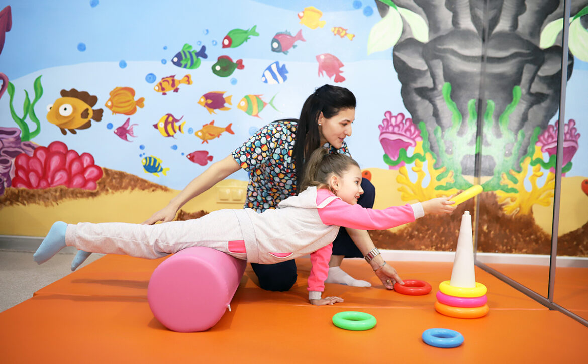
For success in Cerebral Palsy rehabilitation, it is important to determine the problem related to the movement system and to plan and apply physical therapy rehabilitation methods. The purpose of physical therapy and rehabilitation is to help the muscles with excessive contraction to relax, to help the loose muscles that do not contract sufficiently, to increase their balance and coordination abilities and to reveal maximum mobility.

It is very important to start physical therapy and rehabilitation early. Preventing the development of abnormal posture reactions and abnormal contractions, developing normal contraction, preventing the development of joint motion limitation and deformity, giving the child a functional movement in self-care activities such as feeding and dressing are the goals of early treatment. All these activities affect the quality of life positively, albeit with slow development.

Main purpose of treatment is to assist the brain in the formation of the correct motor response by ensuring the integration of all information.

What to Do in Cerebral Palsy Rehabilitation?

Cerebral Palsy treatment is a team work. A team consisting of pediatricians, pediatric neurologists, physical therapy and rehabilitation specialists, physiotherapists, occupational therapists, dieticians and psychologists should closely monitor the child, and if the need for an operation arises, an orthopedic and neurosurgeon should step in. The focus of the team’s work should be on the family. The more conscious, patient and diligent the family is, the more beneficial the baby will be from the medical team.

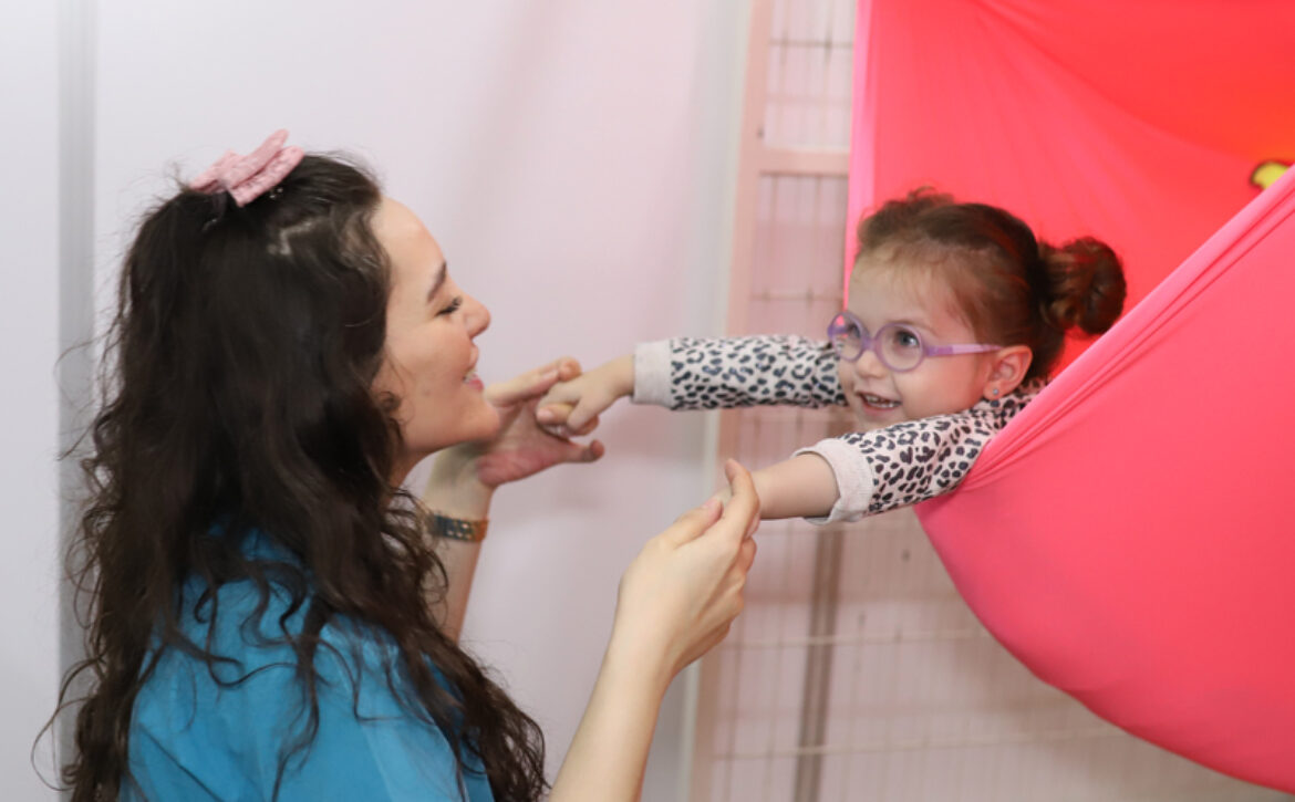
Sensory integration therapy also plays an important role in rehabilitation. Our brain organizes the senses coming from the environment so that we can use our body effectively in our daily life activities and our relationships with our environment. It is either very difficult or impossible to organize this organization in children with cerebral palsy. Sensory integration therapy should be an integral part of rehabilitation to correct this situation caused by impaired sensory input and motor response flow.

In Rommer Physical Therapy and Rehabilitation Center, physical therapy and rehabilitation of our children with Cerebral Palsy is successfully applied. It is applied by experts in Robotic Rehabilitation and Sensory Integration Therapy and this increases the success rate.


25 August 2020 by rommer 0 Comments

Hydrocephalus Rehabilitation

HYDROCEPHALUS REHABILITATION IN CHILDREN

Hydrocephalus is a disease characterized by the accumulation of excessive amount of cerebrospinal fluid (CSF) in the enlarged cerebral ventricles and subarachnoid space.

CSF is a fluid mainly produced in the brain in a structure called the choroid plexus. The rest of the CSF is made by the cerebral tissue.

CSF first passes to the system of cavities in the brain (ventricle) and then to the system under the cerebral membrane (subarachnoid space) and meets the nutritional and fluid needs of the nerve cells in all these regions.

What is Hydrocephalus in Children?

Hydrocephalus means excessive water accumulation in the brain. It is a condition that draws attention with the excessive growth of the head in children. Cerebrospinal fluid (CSF) protects the brain and spinal cord against concussions. In addition, CSF acts as a protective shield against harmful substances in the blood through a mechanism called the blood barrier.

CSF is a fluid that is continuously made and absorbed and removed from the brain and spinal cord. After completing its circulation, it is absorbed into the circulation through the veins. Therefore, any obstacle or abnormality that may occur during production and discharge will cause hydrocephalus. Hydrocephalus occurs as a result of an imbalance between CSF production and absorption in the skull.

What Causes Hydrocephalus in Children?

Hydrocephalus in children is always associated with increased intracranial pressure syndrome (KIBAS).

  • Increase in CSF production in the choroid plexus,
  • Inadequate absorption of CSF,
  • Intracranial pressure will increase due to the increased amount of CSF as a result of congestion, congenital stenosis, etc. that may occur anywhere along the path of the CSF.

The most common cause of Hydrocephalus in children is a disorder in the CSF circulation. As a result, large amounts of CSF accumulate in the cerebral spaces. Less commonly, it occurs as a result of absorption failure. Unless KIBAS increases too much, CSF production amounts will continue to remain within physiological limits.

Enlargement occurs in the brain cavities above the obstruction. As a result, brain tissue damage occurs. If hydrocephalus in infancy develops before the cranial sutures are closed, there will be a significant increase in the circumference of the head. Therefore, intracranial pressure does not increase much and brain tissue damage is relatively less.

If hydrocephalus occurs after the sutures are closed or acutely, a severe KIBAS will occur and the damage to the brain tissue will increase.

  • Hydrocephalus is seen between 0-2 years of age as a result of congenital (most frequently occurring in this way, it may occur only in the form of congenital hydrocephalus or accompany with other syndromes) and as a result of intra-brain hemorrhage.
  • In children and adults, it is seen as a result of brain infections, brain hemorrhage, head trauma and brain tumors.

What are the Symptoms of Hydrocephalus in Children?

Although it varies according to age and person, in the first two months;

  • Head growing more than normal,
  • Thinning of the scalp
  • Clarification of the veins in the head,
  • Vomiting
  • Eyes crossing down,
  • Symptoms such as suffering a seizure are at the forefront.

In addition to these symptoms, different complaints such as nausea, vomiting, visual disturbances, sensory movement disorders will occur in later months and ages.

Vital signs (such as breathing, swallowing, etc.) may be adversely affected as a result of the brain stem being affected. Changes such as slow heart rate, systemic hypertension, respiratory disorders may occur.

How Is Hydrocephalus Diagnosed in Children?

Hydrocephalus can be detected by prenatal ultrasound examination during pregnancy. Serial head circumference measurements should be made after delivery, and ultrasound examination should be prioritized before anterior fontanel closure. Although the lateral ventricles can be seen successfully with ultrasound, the posterior fossa cannot be seen. After the anterior fontanel is closed, imaging methods such as CT or MRI should replace ultrasound in diagnosis.

How Should Hydrocephalus Be Treated in Children?

Hydrocephalus in children cannot be treated with medication. Surgical treatment methods for the underlying cause are preferred. Therefore, the CSF will need to be transferred from the brain to another area in the body by placing a shunt (canal). This operation is performed with a thin catheter. However, since the BOS is produced continuously, the shunt system must be in continuous working condition and open. The shunt can become infected with clogging microbes.

How Should Rehabilitation Be in Children with Hydrocephalus?

  • It is necessary to increase the strength of the muscles in the affected area, and to apply personal therapy.
  • Studies aimed at increasing balance and coordination should be implemented, and walking training should be done by increasing balance with robotic rehabilitation in today’s technology.
  • Ergotherapy is a method that allows the person to adapt to daily life. At the same time, the sensory integration training is given to help the person gain their functions.


25 August 2020 by rommer 0 Comments

Spina Bifida Rehabilitation

What is Spina Bifida?

Spina Bifida; means split or open spine. It is one of the most common congenital diseases. The baby is born with an opening on the back. Different degrees of paralysis may occur depending on the location of the opening. The higher the opening, the more severe the paralysis will be. For this reason, some patients with Spina Bifida need less assistive devices (some do not use them at all), while others may even have to use a wheelchair.

Affected muscles may not only be muscles that allow movement. In the muscles that control urine and stool, they can participate in many patients.

More than 85% of patients with Spina Bifida experience a collection of fluid in the brain (big head). Since the skull bones in babies have not hardened yet, the head grows and tries to reduce the pressure. For this reason, if hydrocephalus is intervened before brain damage occurs, generally the intelligence levels of children with spina Bifida will not be affected.

What Causes Spina Bifida?

Spina Bifida is an anomaly that occurs in the first month of pregnancy in the womb. While the baby’s spine is taking shape, it does not fully close and for this reason, the picture known as “split spine disease” occurs among the people. It is not known exactly what causes it.

In Spina Bifida, nerves and membranes protrude through the open vertebrae, forming a lump on the baby’s back.

First of all, folic acid deficiency is blamed. Therefore, precautions should be taken in women and pregnant women who may become pregnant.

Spina Bifida is more common in caucasians and more common in women. The higher incidence of spina Bifida in the family brings to mind hereditary causes. The risk of giving birth to a baby with spina bifida again in future pregnancies is 15%. Some drugs used in epilepsy, such as valproic acid, are said to increase the risk of giving birth to a baby with Spina Bifida.

How Many Types of Spina Bifida Are There?

Spina Bifida occurs in the first 3 weeks of pregnancy and its incidence is approximately 0.15-0.3%. Although there are three types according to the severity of the disease and the problems it causes, when Spina Bifida is mentioned, myelomeningocele, the most severe type, comes to mind first.

  • Spina Bifida Occulta is the most common and mildest form of the disease. Many people are not even aware of the disease. Often it is noticed as a result of an x-ray had for another purpose. A small portion of the bones in the spine are open and is also called closed spina Bifida. Usually it does not cause any discomfort and does not require surgery.

In rare cases, the spinal cord can be compressed and stretched. As a result, weakness in leg movements and urinary incontinence may occur. This is called tethered spinal cord syndrome.

  • Meningocele is the rarest type of Spina Bifida. Since there are no nerves in this part, which comes out in the form of a sac, it does not cause serious problems. Some babies may have bladder and bowel problems, and rarely hydrocephalus.
  • Myelomeningocele is the most serious and common type of Spina Bifida. The sac that comes out of the spinal bones also contains the spinal nerves. As a result, it may cause some problems such as partial paralysis, walking difficulty, hydrocephalus, urinary / fecal incontinence, advanced renal failure, and scoliosis.

What Are the Symptoms of Spina Bifida?

The involvement of the affected area, spinal cord membranes and nerves are among the factors that will determine the clinical symptoms.

Spina Bifida Occult is usually asymptomatic. Occasionally, visible findings can be found above the defect, such as an abnormal strand of hair, a small depression or a birthmark at the anomaly site.

In the meningocele, the membranes around the spinal cord protrude through the opening in the spine. Because the sac contains only liquid, it can rarely cause hydrocephalus and rarely bladder and bowel problems.

In myelomeningocele, the spinal canal remains open in the lower or middle part of the back, along many vertebrae. Inside this sac are both the membranes surrounding the spinal cord and the spinal cord or nerves. In the later stages of life, serious symptoms such as walking problems, leg deformities and bladder control may occur. Symptoms are characteristic according to the localization in patients. Partial paralysis, walking difficulty and hydrocephalus can be seen.

How Is Spina Bifida Diagnosed?

Spina Bifida can be diagnosed by amniocentesis, ultrasound and blood tests. In the triple test, AFP (alpha feto protein) values are higher than expected, increasing the possibility of Spina Bifida.

The triple test is applied between the 16th and 18th week of pregnancy. Also, Spina Bifida can be detected with a detailed ultrasound test to be performed in these weeks. When Spina Bifida is diagnosed, the skull, spine and other organs should also be examined thoroughly.

How Is Spina Bifida Treated?

If the birth of a baby with Spina Bifida is allowed, it should usually be operated within 36 hours after birth. With the operation, the sac on the back should be closed. In those who develop hydrocephalus, a drainage system called shunt operation should be applied to drain excess fluid from the brain into the blood circulation.

Surgeries are often applied to protect the current situation and prevent further problems. It is not possible for patients to recover completely after the operation. However, even if patients with Spina Bifida cannot become completely healthy, it is possible for them to live independently.

The application of some physiotherapy methods in the postoperative period will be especially beneficial in terms of strengthening the leg and waist muscles.

In rehabilitation, exercises (sitting, balance, walking exercises) should be done to strengthen muscles and prevent joint stiffness. Measures should be taken to prevent pressure wounds, and devices called orthoses to relieve leg weakness should be made. Urinary disorders (urinary incontinence and inadequate ejaculation) may occur in patients. In these cases, urine should be evacuated regularly with a catheter. In terms of kidney health, care should be taken against urine infections and leakage of urine into the kidney. Patients may also develop constipation and precautions should be taken.

Since the event is particularly in the lower spinal cord, there are paralysis of the legs and feet. However, these strokes are often loose. However, there may be defects in the hands due to the occurrence of hydrocephalus. The paralysis can become contractive.

Since these children are prone to obesity, attention should be paid to their diet, and foods such as soda, sugar, chocolate and wafers should not be given.

25 August 2020 by rommer 0 Comments

Muscular Dystrophy Rehabilitation

What is Muscular Dystrophy?

Muscle disease (MD) is the name given to a group of diseases that cause progressive weakness and loss of muscle mass. Muscle diseases often occur at an early age. Although congenital muscular dystrophies start to show symptoms from birth, symptoms in other diseases of this group usually begin after the age of three.

Duchenne Muscular Dystrophy (DMD) is the most common congenital muscle disease. Since the disease is transmitted by the X chromosome, the majority of patients are men. The incidence of this disease, which is 1 / 3.500 in men, is expressed in figures of 1 / million in women. More than 30 muscular dystrophy diseases have been described today. These diseases;

  • Genes that cause disease,
  • Affected muscles,
  • The age at which symptoms first appear,
  • They differ according to the progression age of the disease and are classified accordingly.

What Causes Muscular Dystrophies?

Muscular dystrophies can be genetic or can occur as a result of changes in genes. In Duchenne and Becker muscular dystrophies, there is a deficiency in the synthesis of a protein called dystrophin, which is genetically responsible for muscle nutrition. In this disease, since the dystrophin gene is carried on the X chromosome, it is X-linked and is very common in males.

What are the Symptoms in Muscular Dystrophies?

Most of the symptoms of congenital muscle diseases occur in childhood or adolescence. Congenital muscular dystrophies only start from birth or within the first 2 months and can be seen in both sexes. General symptoms;

  • Walking on toes
  • Having weak muscles, falling frequently
  • Recurrence of muscle cramps
  • Difficulty getting up, climbing stairs, running and jumping
  • Often having a curved spine (scoliosis).

Duchenne muscular dystrophy (DMD), which is the most common among muscular dystrophies, begins to show symptoms at the age of 3-5. Sometimes it may occur at an earlier age with symptoms such as late start to walk, laxity in muscles and falling while walking. Generally, the first finding is the inability to straighten the head from the lying position due to the weakness of the neck muscles. Clumsiness, falling out, inability to play games with peers occur. This situation worries the family. A duck-like, rocking gait occurs as a result of the involvement of the shoulder and hip muscles. Therefore, the first symptoms are difficulties and frequent falls, especially when getting up from the ground, climbing stairs, climbing uphill. Children can often stand up with their hands resting on the ground.

How Do Muscular Dystrophies Progress?

Most of the other congenital muscle diseases, except DMD, which occurs early, start at the age of 10-20. DMD progresses slowly (by creating a decrease in muscle strength) in children and can reach levels that can make them need a wheelchair at the age of 7-12.

These people may need assistance in moving their arms, legs and bodies as a teenager. Most of the patients die in their 20s due to respiratory or cardiac reasons. The muscle breakdown in DMD does not cause any pain in the patient.

How Are Muscular Dystrophies Diagnosed?

The frequent falls of children cause families to consult a doctor.

  • Blood test; CK is very high, genetic tests are applied
  • Muscle biopsy; shows changes in the muscles
  • EMG; indicates disturbances in muscle conduction and contraction
  • Muscle strength and reflex tests help the diagnosis.

How Should the Treatment Be in Muscular Dystrophies?

There is no treatment method for DMD yet. Steroid drugs, some devices and physical therapy can be tried to extend the range of motion in patients.

Supportive methods such as occupational therapy, speech therapy and pulmonary rehabilitation can be used in addition to physical therapy. Exercise should be done in a level that does not create fatigue. In order not to make children fat, it is necessary to stay away from sugary, flour foods and beverages such as cola.

In the treatment, it should be aimed to give the child a happy childhood.

In DMD patients, a special sling should be worn, including the ankles, in order to prevent the ankles from looking downward, especially when lying down.

Mothers known to be carriers of DMD should undergo genetic testing. As a result of genetic tests in new pregnancies, it will be understood to a great extent whether the fetus has DMD.

25 August 2020 by rommer 0 Comments

Spinal Muscular Atrophy (SMA) Rehabilitation

SPINAL MUSCULAR ATROPHY (SMA)

It is a congenital disease of the motor nerves located in the anterior horn section of the spinal cord. The incidence of Spinal Muscular Atrophy (SMA) with genetic inheritance is 1-2 / 10,000.

What is Spinal Muscular Atrophy (SMA)?

In Spinal Muscular Atrophy (SMA), there is no response to the neural stimulation coming from the brain to the anterior horn cells of the spinal cord and enabling the voluntary muscles to work. Because, there is a genetic dysfunction in the motor anterior horn cells. Therefore, since these nerves are not stimulated to the peripheral (peripheral) muscles, the muscles cannot contract voluntarily and myolysis (atrophy) occurs. All arm and leg muscles, trunk and some respiratory muscles are affected by this disease.

What Causes Spinal Muscular Atrophy (SMA)?

There is a mutation in the motor nerve gene (SMA gene) in SMA disease, popularly known as “loose baby syndrome”. In normal healthy people, the motor neuron gene produces a protein critical to the function of the nerves that control our muscles. Thanks to this protein, nerves become strong and carry out orders from the brain. However, if this protein cannot be synthesized, motor neurons are broken down and destroyed. As a result, no signals can be sent to the muscles.

The main feature of all types of SMA is muscle weakness accompanied by muscle wasting.

How DoesSMA Appear?

SMA is examined in 4 different types, considering the age at which it starts and clinical symptoms. The earlier the symptoms of SMA appear, the more severe the course of the disease. SMA type 1 seen under 6 months shows the most severe form of the disease.

  • Type1 SMA is the most severe type, also called hypotonic baby. There is no head control and sitting without support in the sick baby. Even if there is no movement in the arms and legs, patients have eye contact and hearing is normal. Sucking and swallowing difficulties are also a problem in Type 1 SMA, which is one of the most common causes of infant deaths in the world. Frequent respiratory tract infections develop as a result of the respiratory muscles being affected. As a result, lung capacity gradually decreases and sick babies become dependent on breathing apparatus. Their prognosis is not good.
  • Type2 SMA is frequently seen in babies between the ages of 6-18 months. It is observed that the baby whose development is normal suddenly cannot develop and even goes backwards compared to his peers. These patients have head control and can sit alone. However, they cannot move from lying to sitting position. These patients can never stand and walk without support. Respiratory tract infections are frequently seen in these patients, and the risk of developing spinal curvature is also high. It is a relatively slow table.
  • Type 3 SMA is the form that starts in infants older than 18 months. It progresses slower than the other two types. Symptoms such as difficulty in walking, frequent falls, and inability to climb stairs occur due to muscle weakness in the child who was normal until the time of diagnosis. Symptoms can occur at any time until puberty. These patients can stand and walk and their respiratory muscles are not affected. Scoliosis is relatively more common in this group of patients. In advanced ages, they may need a wheelchair.
  • Type4 SMA is the type seen in adulthood and has a good prognosis.

What are the Symptoms ofSMA?

Although the symptoms vary according to the age at which the disease occurs, they may differ from person to person. Although the general symptoms vary according to the type of the disease;

  • Muscle weakness and debility leading to a lack of motor development. The weakness is usually symmetrical, often involving proximal muscles such as the shoulder and hip circumference. The weakness in the leg muscles is more than the arms,
  • They fall behind their peers in terms of motor function,
  • Frequent falls, difficulty climbing stairs, difficulty standing and walking,
  • Shaking hands, tongue twitching,
  • Decrease or loss in deep tendon reflexes (sensation is preserved),
  • Increased risk of aspiration due to feeding difficulties, swallowing and swallowing problems,
  • Failure to control head and waist.

The diagnosis of the disease can be made by genetic analysis and EMG. In genetic analysis, it is diagnosed by detecting the anomaly in the SMN (survivor motor neuron) gene located in the long arm of chromosome 5.

What Are the Treatment Methods of SMA?

Extensive research is ongoing for the treatment of SMA disease. SPINRAZA, the first drug to be approved by the FDA in 2016, is still used in the treatment of children, and losses in patients can be reduced or even stopped with this drug.

In addition to drug treatment, rehabilitation programs to be applied in SMA have very beneficial and positive effects. Purpose in rehabilitation;

  • Preserving the current situation, delaying the deterioration as much as possible,
  • Increasing or maintaining the independence of the child,
  • Reducing the family’s care burden,
  • Prevention of joint deformities,
  • Protection of muscles,
  • Elimination of respiratory complications,
  • The maintenance and provision of roles and life in social life.

SMA disease not only affects the musculoskeletal system in relation to the pathology it creates, but also brings many secondary problems. Treatment principles should be holistic and multidisciplinary, and each patient should be treated individually.

Considering the effects of SMA, orthosis may be required to prevent some problems that may arise in children due to muscle weakness. In addition, it is recommended to use walking devices such as walkers and wheelchairs in accordance with the functional state of the child in order to support independence.

Education of the family about the disease, and the importance of stretching and strengthening exercises to protect joints and muscles should not be forgotten.

The first step in the rehabilitation of SMA disease is to evaluate the patient’s current functional status. It should be repeated every 3-6 months. Because the functional state of the growing child will constantly change. Widespread weakness in muscles despite growth will have adverse effects on both bone and tendons. As a result, growth disorders will occur in the bone. A poorly developed hip joint is an example. Bilateral hip dislocation is a common finding in SMA cases.

The joints of SMA cases are also quite loose. In the early stage of the disease, the range of joint motion may even go beyond what is expected. This condition is a factor that increases spinal scoliosis. Loss of muscle strength, laxity in joints and limitation of movement, and changes in arms and legs due to growth are the main factors affecting functions in patients with SMA.

How is Respiratory Physiotherapy Applied in SMA?

Weakness of respiratory muscles reduces ventilation of the lungs in patients with SMA. This situation leads to frequent lung infections and diseases. As a result of the weakness of the cough-related muscles, secretions become difficult to expel. Children with widespread muscle weakness will also have an increased risk of aspiration.

Small oral foods, the use of straws, keeping the head upright during feeding and chin support, less and frequent feeding are practices that will help prevent the risk of aspiration.

From an early stage, it is important to develop muscle strength, maintain the flexibility of the rib cage, increase the range of motion and improve physical functions. Appropriate positions should be shown by physiotherapists for comfortable breathing.

Positioning and various drainage methods will be useful in the disposal of lung secretions. Appropriate postural drainage measures should definitely be taken to prevent aspiration in patients and the family should be educated on this issue.


25 August 2020 by rommer 0 Comments

Risky Baby (Premature) Rehabilitation

WHAT IS A RISKED BABY?

Babies who may show growth retardation due to problems experienced during pregnancy, delivery or postpartum are called risky baby. In general, the group of risky babies can include premature babies (premature) babies, term babies with a low birth weight and babies who need to be looked after in the neonatal intensive care unit after birth.

Which babies are called premature babies?

The baby born before 37 weeks is called premature baby. Babies born between 34-36 weeks are classified as late preterm, babies born between 32-33 weeks are classified as moderate preterm, babies born between 28-31 weeks are classified as very preterm and babies born before 28 weeks are classified as extremely preterm.

What does low birth weight mean?

A birth weight below 2500 grams is a low birth weight. If this measurement is less than 1500 grams, it is classified as very low, and less than 1000 grams as extremely low birth weight babies.

What are the problems that can be seen in risky babies?

In the neonatal period, brain involvement due to lack of oxygen (hypoxic ischemic encephalopathy), neonatal breathing difficulties (respiratory distress), jaundice (hyperbiluribinemia), eye problems due to incomplete development of the vascularization in the eye (retinopathy of prematurity) can be seen. In addition, 5-15% of premature babies may develop permanent neurological conditions such as cerebral palsy.
Risky babies are at risk in terms of delay in motor, sensory, cognitive and social development stages. Weakness in the body muscles, delay in movements such as turning, sitting, crawling, walking, deficiency in sucking-swallowing skills in the early period, and delayed speech in the future can be seen. Risky babies may have difficulty in calming themselves, they may cry frequently and avoid moving. They may have sensory problems such as excessive mobility or needing too much stimulation.

Who should follow risky babies?

The risk level of babies is determined according to the above-mentioned definitions and according to this degree, a neonatologist, developmental pediatrician, pediatric neurologist, special education specialist, occupational therapist, speech therapist and child physiotherapist can be in the team.

How is the neuromotor development of babies at risk assessed?
In our institution, the neuromotor development of babies at risk is done by physiotherapists who know the typical development of babies very well and can observe and analyze the baby’s movements. If the spontaneous movements of babies, position transitions (for example; turning over the back), movement strategies (crawling, on the butt, crawling, etc.), reflexes and muscle tone are examined in detail, the typical development of the baby or deviations from typical development can be learned. In the light of these evaluations, a consensus is reached with the family and the early intervention program of the risky baby is started.

What is an early intervention program?
Early intervention program includes physiotherapy approaches starting as early as possible (as soon as possible) and up to 24 months. Depending on the risk level and condition of the child, the physiotherapy program can be continued for longer periods in the presence of neurological effects until school age.

Purpose of physiotherapy programs is using the self-renewal (plasticity) feature of the brain, the acquisition of typical developmental movements and bringing the child to the most independent level possible. In this respect, the first 2-3 years of life are very valuable because brain and nerve development is very rapid in these years, so early intervention will increase the success of the treatment.

Bobath (neurodevelopmental) therapy is the most widely used approach worldwide to support motor and sensory development in infants. Purpose in Bobath concept is to give children with neurodevelopmental disorders the highest level of functionality possible. For this purpose, bobath therapists create an intervention program that includes objectives such as providing the necessary environmental adaptations for the baby, increasing the communication between the baby and the family, including the family in the therapy, and teaching the therapy to spread throughout the day, as well as specialized therapies for the baby. The baby is constantly evaluated, this program is arranged at intervals according to his needs and the effectiveness of the treatment is observed.

How can I access the assessment and early intervention program for babies at risk?
In the bobath therapy unit in our institution, an assessment and early intervention program is applied in risky babies. Risky babies are evaluated in the bobath therapy unit and their development is supported with a physiotherapy program specific to the baby.


25 August 2020 by rommer 0 Comments

Brachial Plexus Rehabilitation

BRACIAL PLEXUS INJURY

The nerve roots originating from the spinal cord in the neck region, traveling as 3 large branches, join in the armpit space and form a large nerve network. Brachial plexus injury occurs as a result of the lesions of the nerve network in this region.

Nerves from the Brachial Plexus to the arm exit from the spinal cord. This neural network includes 4 large cervical (neck) nerve roots (C5-C8) and first dorsal (thoracic) nerve root (T1). These roots unite and divide into 3 main branches. The branches from the C5-C6 roots form the upper branches, the branches from the C7 roots form the middle, and the branches from the C8-T1 roots form the lower large branches.

Each major branch is divided into sections and moves the muscles (flexors), some of which generally bend the arm and raise it up (flexor), and other parts to the muscles that extend the arm and lower it (extensor).

What is Brachial Plexus Injury?

Brachial Plexus injury is the development of paralysis at varying degrees in the arm muscles stimulated by this nerve as a result of damage to the nerve network (Brachial Plexus) that goes from the brain to the arm through the spinal cord. Although it varies according to the affected nerve roots, paralysis starting from the arm muscles can reach the fingers of the hand.

What Causes Brachial Plexus Injury?

Paralysis that develops in varying degrees as a result of the Brachial Plexus injury, is a condition that starts in the neonatal period and continues throughout childhood, and requires long-term follow-up and rehabilitation. Most of these injuries occur during birth. However, Brachial Plexus injury has also been reported in deliveries by cesarean section. Mostly;

  • High birth weight (especially over 4 kg)
  • Difficult and prolonged birth,
  • Use of auxiliary tools such as vacuum and forceps during birth,
  • Shoulder or breech presentation,
  • And many other reasons such as structural strictures in the mother are blamed for Brachial Plexus injuries.
  • Traumas in the nerves;
  • Avulsion type (ruptured from the peripheral synoromuric)
  • In the form of neuropraxia (edema around the nerve) or
  • Axonotmesis (nerve sheath is normal, only axon damage is present).

Nerve rupture is irreversible. Regeneration is possible in other types of injuries.

What are the Symptoms of Brachial Plexus Injury?

Since the Brachial Plexus takes nerve fibers from the roots between C5 and T1, the paralysis that will occur will vary according to the level of the nerve involved. Although it is generally examined under three headings, it can show transition to each other.

  • Upper trunk (nerve root) involvement is called Erb-Duchenne type paralysis and it is the most common type. Babies with this type of injury cannot move their shoulders and have difficulty extending and rotating their arms. If C7 involvement occurs, fingers curl in the position of asking for tips. If C4 involvement takes part, the phrenic nerve is affected and related symptoms also occur.
  • Lower trunk (nerve root) involvement is called Klumpke’s palsy. Isolated lower trunk injury is rare. Arm, shoulder and elbow movements are good in classical condition. However, hand claw deformity is observed. As a result of this type of damage, weakness occurs in the hand and wrist, but shoulder and arm movements are generally preserved. In lower trunk involvement, the sympathetic cervical chain is rarely affected and Horner syndrome occurs.
  • The third type, called complete involvement, is also called Erb-Klumpke. The upper, middle and lower trunks (nerve roots) of the Brachial Plexus are affected to varying degrees. Although this varies depending on the degree of involvement in babies, it can be observed in complete motor and sensory paralysis. Weakness occurs in shoulder, arm, hand and wrist movements. When fully developed, areflexia and complete loss of sensation are observed. 65% of these patients have Horner syndrome. (Horner syndrome is usually characterized by miosis, ptosis, enophthalmos and loss of sweating in the half of the affected face as a result of involvement of the lower turuncus, cervical sympathetic chains.)

In 80% of the cases, spontaneous recovery occurs in the first 3 months. Although it varies according to the severity of the injury and the degree of involvement, various clinical pictures will occur.

How Is Brachial Plexus Injury Diagnosed?

Besides the clinical findings of the patient, radiological high resolution MR imaging technique is one of the best diagnostic options. Thanks to this test, the soft tissue will be seen much better. Fractures in the bone structure such as the clavicle and humerus can be detected with direct radiological studies.

EMG is a very valuable diagnostic method for understanding nerve and muscle damage. It should be applied two to three weeks after injuries. It will give information about the localization and degree of the injury.

How Is Brachial Plexus Injury Treated?

After the diagnosis, a team approach should be applied to achieve the best result and the patient should be evaluated accordingly.  In the treatment team, there should be especially family, children, physical therapy, orthopedics and neurology specialists and physiotherapists, and the treatment methods to be applied should be taken by common decision.

There may be many different treatment approaches ranging from conservative treatment to surgical interventions. Conservative treatment methods such as physiotherapy and occupational therapy should be tried initially. If success is achieved, treatment should be continued until neurological functions return completely to normal or a plateau in functions is reached. The aim of rehabilitation in conservative treatment is to prevent the development of contractures in the periods where healing is expected by preserving passive joint movement, flexibility of joints and muscle strength.

  • Reducing joint stiffness in the affected arm
  • Informing the family about the positioning of the affected arm
  • Ensuring active use of the affected arm
  • Increasing and strengthening the baby’s arm movements.

Surgical indications and timing issues in Brachial Plexus injury have always been controversial. It may differ considerably according to many centers and surgeons.


25 August 2020 by rommer 0 Comments

Bobath Rehabilitation

BOBATH THERAPY IN CHILDREN

What is Bobath Therapy?

Bobath Therapy is one of the most preferred physiotherapy methods in the world, which is used in the rehabilitation of children and adults.  It can be applied regardless of the presence of accompanying problems especially in Cerebral Palsy. It is also widely used in other conditions that cause movement disorders and sensory and motor developmental delay.

The foundations of Bobath therapy were laid in the 1940s by physiotherapist Berta Bobath and his wife, neurophysiologist Karol Bobath. Today, it is a dynamic treatment method that is still updated with scientific studies.

What is the purpose of Bobath Therapy?

Bobath therapy is specially planned for each child. The main purpose of the therapy is to increase the functional capacity of the child, to diversify the activities that he / she can do and to encourage him / her in this direction. In this way, the child’s adaptation to his environment will increase, his / her communication with people will be strengthened and he / she will reach the maximum level of independence that he / she can have in every aspect of his / her life.

What is the basis of Bobath Therapy?
Bobath therapy mainly focuses on the central nervous system. The self-restructuring (neuroplasticity) feature of the brain is used.

Rather than teaching a movement such as sitting, walking, etc, a very good assessment should be made of why the child is unable to do that movement. In order for this movement to be applied, a suitable environment should be prepared and action should be taken directly.

What are the techniques used in Bobath Therapy?
Since Bobath therapy is specific to the individual, the degree of central nervous system involvement and motor dysfunction in the child are analyzed first. The functional boundaries of the child are determined. As a result of this analysis, short and long term goals in therapy are determined with the family.  In the therapy part, special therapeutic grips, positioning, stimulation and facilitation techniques are used. With these techniques, it should be aimed to regulate abnormal muscle tone, to give the child a normal sense of movement, and to reveal the correct movement in the child.


What distinguishes Bobath Therapy from other methods?
Active participation of the child in Bobath therapy is one of the strengths of the concept. Conducting the sessions in play activities in an enriched environment ensures the participation of the child and increases the motivation required to make the movement. Children learn by playing.

Another strength of the therapy is that it is family oriented. Home and school arrangements should be suitable for the child, the reasons for this situation should be explained to the family in detail. In line with the purposes of the sessions, the family should be offered the activities they can do at home. The treatment continues with orthotic supports and assistive devices aimed at increasing the independence of the child and protecting the body alignment. Evaluations should be continuous in subsequent controls.

Who applies the Bobath therapy?

Bobath therapy is performed by physiotherapists, occupational therapists or speech-language therapists who are specialized in pediatrics for children and who have successfully completed their education and received the title of “Bobath therapist”.

How can I get Bobath Therapy?

If your child has problems such as neurological and developmental retardation and difficulty in moving, you can apply to our Rommer International Physical Therapy and Rehabilitation Center to benefit from Bobath Therapy.  In our center, evaluation and Bobath therapy support is provided for babies and children by Bobath physiotherapists.


25 August 2020 by rommer 0 Comments

Yoga Therapy for Children with Disabilities

WHAT ARE THE BENEFITS OF YOGA FOR CHILDREN WITH DISABILITIES

What is Yoga?
Yoga is a science-based spiritual discipline focused on creating harmony between mind and body. In rehabilitation with yoga techniques; balanced yoga poses (asanas) combined to increase body awareness and strength, special breathing (chanting) exercises that improve focus and concentration, improving vocal and speaking skills, eye exercises and relaxation techniques, breathing techniques that strengthen the lungs and allow the whole body to receive oxygen are used.

What are the General Benefits of Yoga for Children?
Rehabilitation with yoga techniques for children is a set of movements consisting of individual and paired exercises, in which classical yoga postures are transformed into appropriate standards for children, combined with fun, discipline, self-confidence and sense of responsibility.

Yoga aims to teach children to use their imaginations with postures, to breathe properly and how to relax their bodies.
Yoga poses (asanas) can help any child with indigestion problems, sleep disorders, stress problems, balance and coordination problems, sensory processing problems or joint and muscle problems. Using their imagination improves their learning skills and enables them to direct their energies in the right direction. However, it can help strengthen muscles, increase focus and creativity, and strengthen the immune system.
Rehabilitation with yoga techniques for children includes breathing exercises, games and stories as well as entertaining yoga postures. The inclusion of games and stories also increases the child’s self-confidence, positive thinking and easier expression of emotions.

The aim of these studies is to balance the body, mind and spirit harmony, as in adult yoga, and to help the child to continue living with this balance.

For Which Disabled Children Is Yoga Beneficial?
Yoga can be applied to children with emotional and behavioral disorders, Down syndrome, cerebral palsy (CP), dyslexia, microcephaly, autism spectrum disorder. It is equally safe and effective as children, even in infants.

What are the Benefits of Yoga for Children with Down Syndrome?
Yoga poses (asanas) help children with Down syndrome stretch their bodies, increase muscle tone and strengthen them. Asanas benefit internal organs and help balance and rejuvenate the endocrine glands.
Children with Down syndrome who practice yoga are weaker and more flexible, while those who do not get enough physical exercise tend to gain weight as they get older. Along with yoga breathing exercises, which have a beneficial effect on the central nervous system, it helps the development of body awareness, concentration and memory. These are very important skills for any child with a developmental disability.

What are the Benefits of Yoga in Cerebral Palsy?
The practice of yoga poses (asanas) followed by deep relaxation can help significantly reduce the high muscle tone that is characteristic of most children with cerebral palsy. Asanas relax the muscles and tendons, relieving tension in the muscles and around the joints. Asanas relax the body; they also strengthen areas of the body with low muscle tone.
One of the most important aspects of asana practice for children with cerebral palsy is stretching and aligning the spine. As a result, the child can develop greater range of motion and coordination as well as greater independence.

What are the Benefits of Yoga in Microcephaly?
Yoga poses (asanas) are an effective way to increase blood circulation and benefit internal organs for less physically active children. Breathing exercises (pranayama) in yoga strengthen the immune system and increase the oxygenation of the blood, which aids in cell repair. By increasing the amount of oxygen in the blood, it sends more oxygen to the brain, which helps strengthen the central nervous system.

Yoga helps children with microcephaly develop body awareness and improve their cognitive and motor skills.

What are the Benefits of Yoga in Autism?
The first step in doing yoga with a child with autism is to establish a strong bond with the child. To do this, the therapist enters the child’s world and establishes a relationship with the child at his or her level. Once the therapist is successful in this relationship, the child gains their full trust. Massage, music, dance, nursery rhymes and stories are some of the different techniques a therapist can use to connect with the child.

As trust and friendship builds between the child and the therapist, the therapist begins to teach the child some yoga poses (asanas) and breathing exercises (pranayama). Once the child is familiar with the initial poses, the therapist can add more asanas and deep relaxation techniques to their work.

The combination of asanas, pranayama and deep relaxation will strengthen the child’s nervous system, increase overall health, and help develop body awareness and concentration. Yoga therapy helps children with autism gain motor, communication and social skills by creating optimum physiological and psychological integrity.

What are the Benefits of Yoga for Attention Deficit Hyperactivity Disorder (ADHD)?
Yoga uses postures (asanas), breathing exercises (pranayama) and deep relaxation techniques to strengthen the central nervous system. Children with ADHD often have learning difficulties due to hyperactivity and distraction.
As a result of yoga, children with Attention Deficit Hyperactivity Disorder (ADHD) focus better at school, their behavior problems are reduced, and anxiety begins to decrease in children with high anxiety levels. A significant decrease in hyperactivity behaviors and a significant increase in concentration are achieved. Students’ ability to focus on a subject and maintain their attention on a task increases significantly after practicing yoga.

With the right breathing techniques, it increases the ability to calm the brain and balance the left and right hemispheres (lobe) of the brain.

What are the Benefits of Yoga for Children with Dyslexia?
The main goal of a child with learning disabilities is to strengthen all areas of the child’s development. Yoga offers an effective therapeutic alternative for children with learning difficulties.
Breathing exercises (pranayama) in yoga stimulate the central nervous system and strengthen the immune system. Along with yoga poses (asanas) and deep relaxation, pranayama makes it possible for the child to develop body awareness, balance, memory and concentration.

Yoga for children with dyslexia uses yogic eye exercises that strengthen the optic nerve, relax facial muscles and stimulate various centers of the brain. These exercises improve the eye’s word recognition and visual perception skills, focusing ability.